An Inside Look
High-tech Imaging
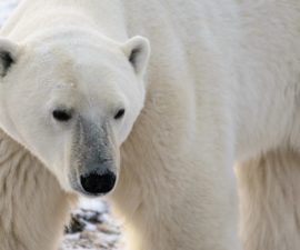
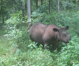
From a tiny poison frog to a formidable two-ton rhinoceros to a finely boned fruit bat to an endangered Mang Mountain pitviper, technology plays a role in ensuring the animals at the San Diego Zoo and Safari Park are thriving.
BY Karyl Carmignani
Photography by Ken Bohn
When an animal at the Zoo or Park presents symptoms that something’s not quite right, exotic animal veterinarians work with wildlife care specialists and veterinary technicians to solve the mystery. It’s not unlike putting together a jigsaw puzzle. High-tech tools like radiographs (also called x-rays), CT scans, and MRIs complement physical exams, blood samples, and expert observation. Together, the team gathers pieces of the puzzle to make a diagnosis.
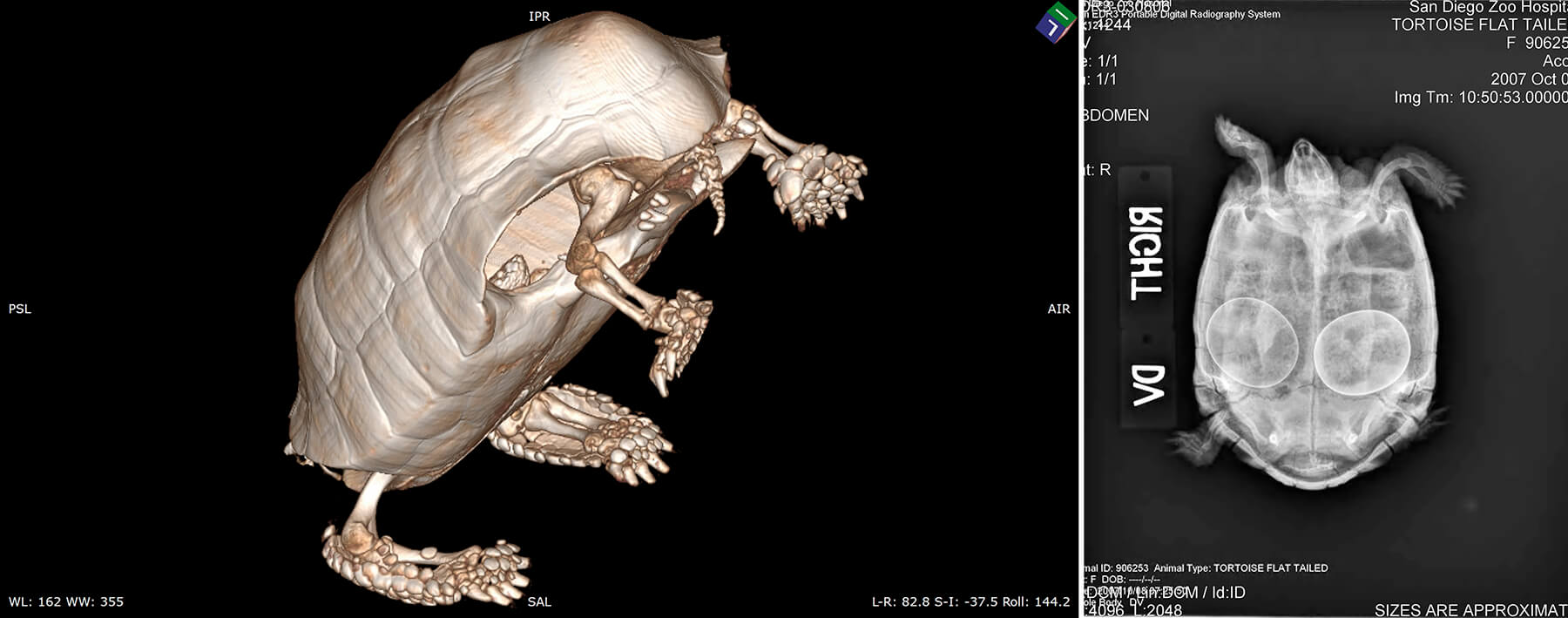
CLOSER LOOK
An MRI (left) and a radiograph (right) provide detailed views of a tortoise.
Radiographs are detailed images of bones and soft tissues. (“’X-rays’ are actually the type of radiation used to produce radiographs,” explained Meg Sutherland-Smith, DVM. “Despite its entrenchment in popular language, the correct name for the images is radiographs, not x-rays.”) Bones and calcium appear white, or “radiopaque,” while the less dense, dark gray areas are said to be “radiolucent.”
While a radiograph image is a still shot, ultrasound technology allows us to see organs in motion. Ultrasound can show blood flow or leaky heart valves, or help guide experts in inserting a tiny biopsy needle into a cyst or tumor.
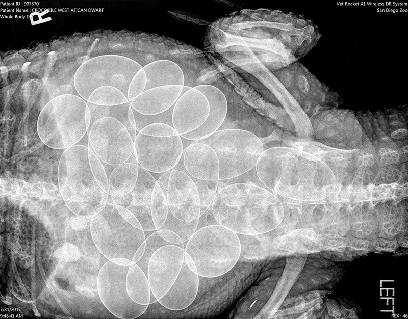
COUNTING YOUR EGGS BEFORE THEY HATCH
A radiograph of a West African dwarf crocodile shows that she’s carrying a clutch of eggs.
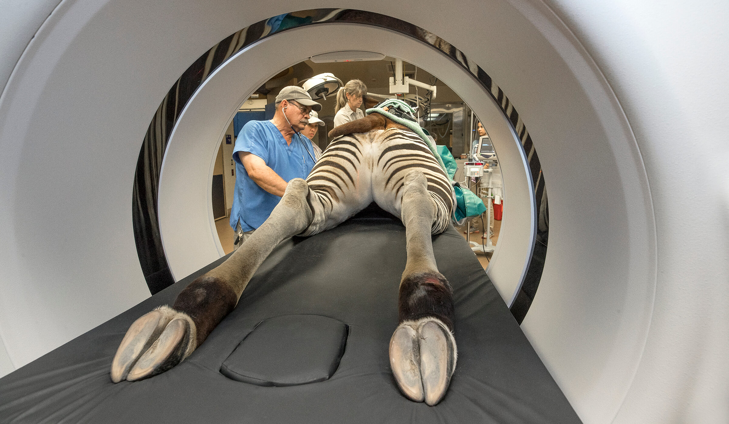
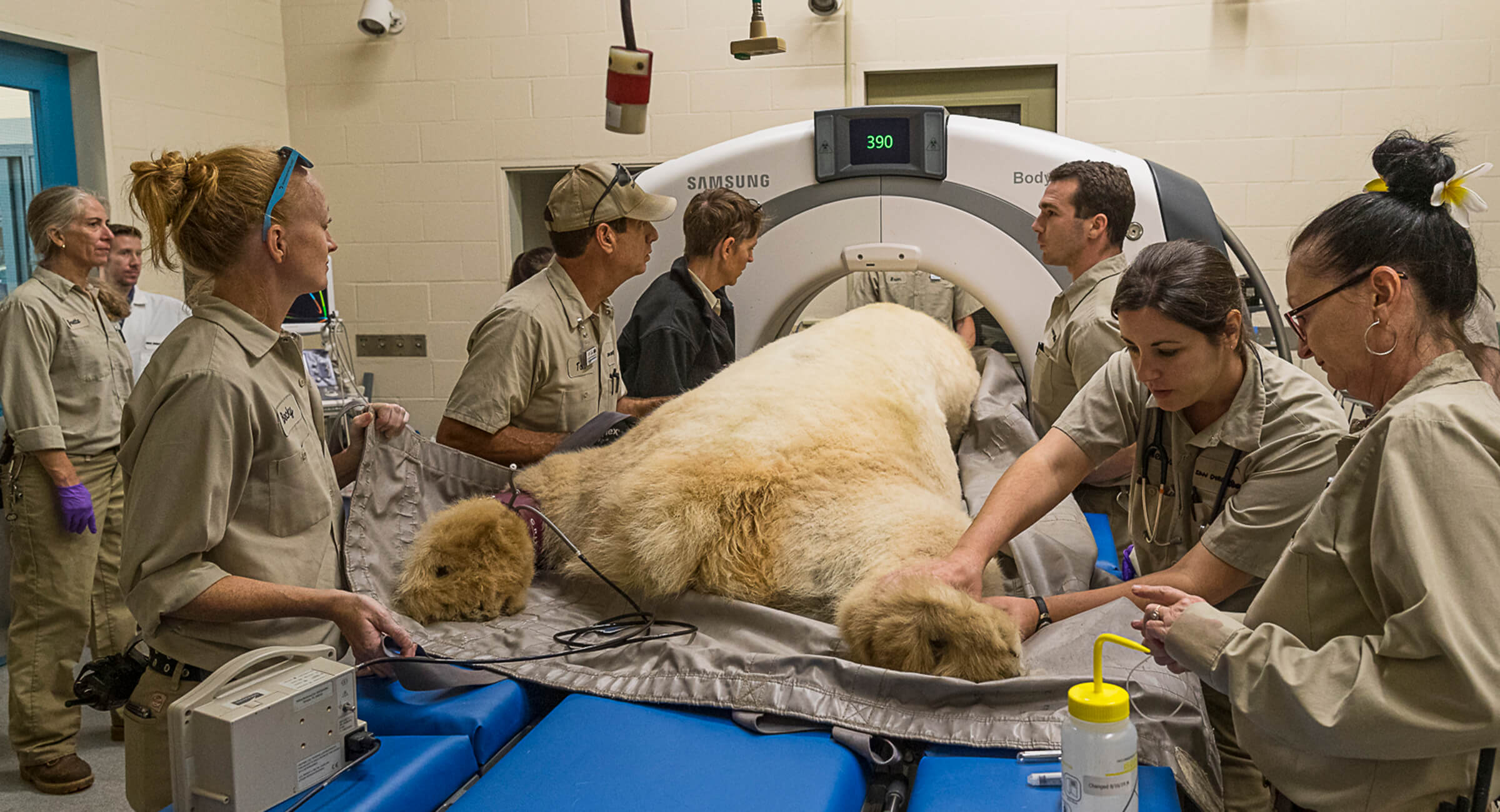
CT or CAT scan stands for computed tomography and uses radiation with computer technology to produce a detailed, cross-section image of the body—revealing intricate details of bones, organs, blood vessels, and soft tissue that cannot be seen in conventional radiographs. CT scans help veterinarians detect, examine, and diagnose bone injuries, lung issues, and various cancers. As the procedure takes less than five minutes, it is often helpful in emergency situations.
MRI, or magnetic resonance imaging, uses magnetic fields and a sophisticated computer to take high-resolution pictures of bones and soft tissue while the patient lies still in a tube-shaped scanner—a procedure that takes 45 to 90 minutes. An MRI can detect soft-tissue injury or detect tumors.
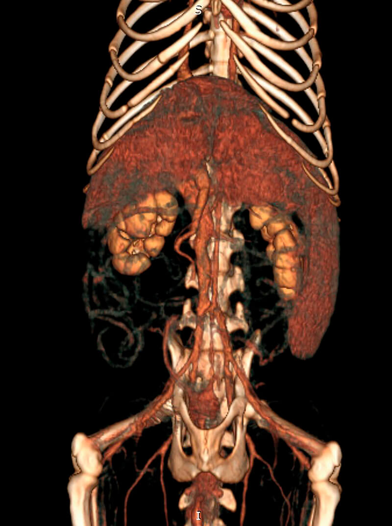
THE INNER OTTER
An MRI captures a 3D image of the skeleton and organs of an otter.
Baseline Basics
Care starts even before a new animal enters the Zoo or Safari Park; a quarantine period ensures that new animals do not bring diseases with them into the Zoo or Park population. Each quarantined animal gets a complete medical exam, including radiographs, so the hospital has baseline medical information about each animal. Sometimes this routine procedure can be lifesaving. For instance, the radiograph of an African spotted-necked otter revealed a large mass next to its heart. Because it was discovered early, veterinarians were able to successfully remove it.
Hold Still
To ensure the images are clear, many animals that require high-tech imaging are anesthetized. But some animals, like polar bears, are trained to participate in their ultrasound procedure. For safety reasons, vets stay outside the enclosure and use a probe extender to roll over the animal’s abdomen while watching the images on a screen. After a bonobo injured her hand, she was trained to place the sore extremity on a flat surface and hold still while a radiograph was taken to ensure it was healing properly.

CHECK IT OUT
A Parson’s chameleon eyes a radiograph of its tail.
Smaller animals like frogs, lizards, hedgehogs, and some birds are tricky to get in detail, so the veterinary team puts the little guys on a mammography machine. It takes highly detailed images of bone, muscle, tissue, and organs, while exposing the animal to less radiation. On the other end of the size scale, elephant foot health is monitored using a portable radiograph machine; the mighty giants are trained to place a foot on a reinforced platform and hold still while the radiograph is taken. Their willing participation is rewarded with delicious treats.

DENTAL EXAM
Veterinarians regularly monitor the teeth of all the Zoo’s koalas.
Sometimes imaging equipment goes out into the field. A portable ultrasound unit provides high-quality scanned images even under glaring sunlight conditions, as a veterinarian dons a head plate with goggles. On St. Bees Island in Australia, a hand-held, battery-operated, dental x-ray unit with sharp, reusable plates documents the koalas’ teeth. (Back at home, veterinarians monitor the bone and teeth health of koalas at the Zoo, too.)

LEARNING FROM IMAGING
Radiographs show (left to right) a boa’s most recent meal, a wallaby’s abdominal organs, and the skeleton of a Pacific horned owl.
Oldies but Goodies
Our radiograph technology went digital in 2005. Prior to that, the veterinary team took films, and these archived radiographs are catalogued in a film library, which is about the size of a walk-in closet, with floor-to-ceiling shelves full of alphabetized manila folders. Given the huge variation among species, it’s a great resource to have, and some of the older animals in the collection have images of their younger skeletons on file. For instance, if an animal had a strange-looking larynx, a vet could go back through the individual animal’s archived images to see what its normal baseline looks like. The scores of aged folders, lined up like sentinels, serve as a reminder of how things were done “old school.” The staff hopes to one day secure funding to digitize this x-ray film library.

BUN IN THE OVEN
A radiograph of a female Allen’s swamp monkey shows her developing offspring.
Software and digital imaging enables staff to amplify details by increasing contrast, switching dark and light areas, and measuring and comparing images, and they make it possible to bring more diagnostic tools out into the field. The images can also reveal some surprises, like a dollar and some change in the belly of a sea lion or a metal bolt in the gizzard of a bird. Some radiograph surprises are happy, like a gently sketched outline of a growing baby inside a monkey. Whatever the images reveal, using modern technology means we can put together a picture of animal health–and keep our animals thriving.